Bioinformatics ReviewBioinformatics Review
Notification Show More
Font ResizerAa
  •  Home
  • Docking
  • MD Simulation
  • Tools
  • More Topics
    • Softwares
    • Sequence Analysis
    • Algorithms
    • Bioinformatics Programming
    • Bioinformatics Research Updates
    • Drug Discovery
    • Phylogenetics
    • Structural Bioinformatics
    • Editorials
    • Tips & Tricks
    • Bioinformatics News
    • Featured
    • Genomics
    • Bioinformatics Infographics
  • Community
    • BiR-Research Group
    • Community Q&A
    • Ask a question
    • Join Telegram Channel
    • Join Facebook Group
    • Join Reddit Group
    • Subscription Options
    • Become a Patron
    • Write for us
  • About Us
    • About BiR
    • BiR Scope
    • The Team
    • Guidelines for Research Collaboration
    • Feedback
    • Contact Us
    • Recent @ BiR
  • Subscription
  • Account
    • Visit Dashboard
    • Login
Font ResizerAa
Bioinformatics ReviewBioinformatics Review
Search
Have an existing account? Sign In
Follow US
Protein VisualizationTools

How to find disulfides in protein structure using Pymol?

Tariq Abdullah
Last updated: January 14, 2021 3:48 pm
Tariq Abdullah
Share
1 Min Read
find disulfides in protein structure using pymol
SHARE

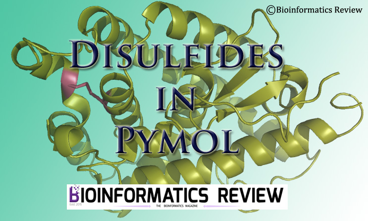
As shown in previous articles, Pymol [1] has several functions. In this article, we will show how to look for disulfide in protein crystal structures using Pymol.

Follow these steps:

  1. Open Pymol by typing pymol in a terminal in Ubuntu and in Windows, by double-clicking the shortcut or by searching.
  2. Go to File --> Open. Select a PDB structure to analyze.
  3. Set the color of the structure and background as you wish.
  4. Go to the right panel, click on the “S” column, it will show many functions.
  5. Select disulfides --> sticks. You can select any representation as you want. Other options include lines and spheres.
  6. If there are any disulfides present in the structure, it will display on the screen.

Make sure you haven’t made sticks/lines/spheres hidden from the “all” row in the top right panel.


Reference

  1. The PyMOL Molecular Graphics System, Version 1.2r3pre, Schrödinger, LLC.
TAGGED:disulfides in pymolprotein visualizationPymol
Share This Article
Facebook Copy Link Print
ByTariq Abdullah
Tariq is founder of Bioinformatics Review and Lead Developer at IQL Technologies. His areas of expertise include algorithm design, phylogenetics, MicroArray, Plant Systematics, and genome data analysis. If you have questions, reach out to him via his homepage.
Leave a Comment

Leave a Reply Cancel reply

You must be logged in to post a comment.

How to visualize a 3D structure using Py3Dmol?
Bioinformatics Programming Cheminformatics GitHub Python
ai tools vs traditional tools in bioinformatics
AI Tools vs Traditional Tools in Bioinformatics- Which one to select?
Algorithms Artificial Intelligence Machine Learning Software Tools
AI vs Physics in Molecular Docking
AI vs Physics in Molecular Docking: Towards Faster and More Accurate Pose Prediction
Artificial Intelligence Drug Discovery Machine Learning
10 years of Bioinformatics Review: From a Blog to a Bioinformatics Knowledge Hub!
Editorial

You Might Also Like

Pymol basic functions- part II
Protein VisualizationToolsVideo Tutorial

Video Tutorial: Pymol Basic Functions- Part II

November 23, 2024
PyMol: Uses & Applications
SoftwareTools

PyMol: Uses & Applications

March 1, 2022
SoftwareTools

Installing MODELLER on Linux/Ubuntu

November 7, 2021
DockingDrug DiscoverySoftwareToolsVideo Tutorial

Video Tutorial: How to perform docking using Autodock Vina

November 23, 2024
Copyright 2024 IQL Technologies
  • Journal
  • Customer Support
  • Contact Us
  • FAQs
  • Terms of Use
  • Privacy Policy
  • Cookie Policy
  • Sitemap
Welcome Back!

Sign in to your account

Username or Email Address
Password

Lost your password?

Not a member? Sign Up