Bioinformatics ReviewBioinformatics Review
Notification Show More
Font ResizerAa
  •  Home
  • Docking
  • MD Simulation
  • Tools
  • More Topics
    • Softwares
    • Sequence Analysis
    • Algorithms
    • Bioinformatics Programming
    • Bioinformatics Research Updates
    • Drug Discovery
    • Phylogenetics
    • Structural Bioinformatics
    • Editorials
    • Tips & Tricks
    • Bioinformatics News
    • Featured
    • Genomics
    • Bioinformatics Infographics
  • Community
    • BiR-Research Group
    • Community Q&A
    • Ask a question
    • Join Telegram Channel
    • Join Facebook Group
    • Join Reddit Group
    • Subscription Options
    • Become a Patron
    • Write for us
  • About Us
    • About BiR
    • BiR Scope
    • The Team
    • Guidelines for Research Collaboration
    • Feedback
    • Contact Us
    • Recent @ BiR
  • Subscription
  • Account
    • Visit Dashboard
    • Login
Font ResizerAa
Bioinformatics ReviewBioinformatics Review
Search
Have an existing account? Sign In
Follow US
SoftwareStructural BioinformaticsTools

How to generate electron density map using Pymol?

Dr. Muniba Faiza
Last updated: November 4, 2022 9:17 am
Dr. Muniba Faiza
Share
3 Min Read
How to generate electron density map using Pymol?
SHARE

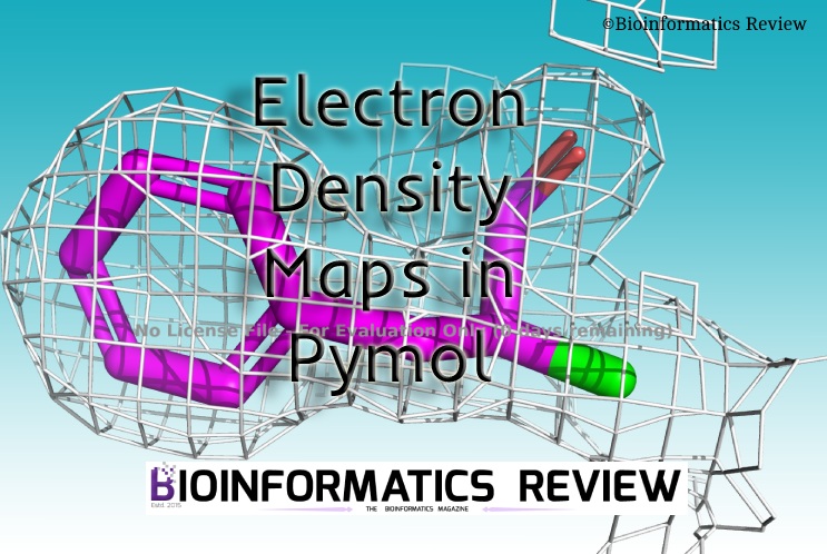
Electron density maps are available for most of the protein structures in PDB. Therefore, in this article, we are using PDB to generate electron density maps in Pymol.

Contents
Electron Density Map of whole proteinElectron Density Map of selected residueReferences

Electron Density Map of whole protein

Follow these steps to generate an electron density map of the whole protein structure:

  • Open Pymol. We will use the command line.
  • Set the view. This is optional, if you don’t want to set the view then skip to the next step.
    > set orthoscopic
    > set line_width, 5
  • Download the structure and electron density data from PDB.
    > fetch 1db1, async=0
    > fetch 1db1, type=2fofc, async=0
  • Now, create the map at 2.5-angstrom carve and map level of 1.06.
    > isomesh 1db1_map,1db1_2fofc, 1.06, poly, carve=2.5
    You can adjust the map level. But if you increase this number then it will become shorter on display.
  • Select a residue to focus on.
    > orient i. 279
    It will zoom in on the selected residue.
  • You can set the colors as well.
    > color magenta, e. C
    > color yellow, e. N

Electron Density Map of selected residue

Follow these steps to generate an electron density map of selected residue:

  • Open Pymol. We will use the command line.
  • Set the view. This is optional, if you don’t want to set the view then skip to the next step.
    > set orthoscopic
    > set line_width, 5
  • Download the structure and electron density data from PDB.
    > fetch 1db1, async=0
    > fetch 1db1, type=2fofc, async=0
  • Select a residue and extract it.
    For example, select residue 279 –> right side panel –> ‘sele‘ –> ‘A‘ –> ‘extract object‘.
    Rename the object if you want. Here, we are using the default name (obj01).
  • Hide the rest of the protein. This step is optional, if you wish to display the rest of the protein then skip to the next step.
    Go to –> Right side panel –> ‘1db1‘ –> ‘H‘ –> ‘hide everything’. Or just simply click on the name of the structure (1db1).
  • Now, create the map at 2.5-angstrom carve.
    > isomesh mesh_ligand_carved, 1db1_2fofc, selection=(obj01), carve=2.5
  • Select a residue to focus on.
    > orient i. 279
    It will zoom in on the selected residue.
  • You can set the colors as well.
    > color magenta, e. C
    > color yellow, e. N

References

  1. The PyMOL Molecular Graphics System, Version 1.2r3pre, Schrödinger, LLC.
TAGGED:electron density mapspdbPymol
Share This Article
Facebook Copy Link Print
ByDr. Muniba Faiza
Follow:
Dr. Muniba is a Bioinformatician based in New Delhi, India. She has completed her PhD in Bioinformatics from South China University of Technology, Guangzhou, China. She has cutting edge knowledge of bioinformatics tools, algorithms, and drug designing. When she is not reading she is found enjoying with the family. Know more about Muniba
Leave a Comment

Leave a Reply Cancel reply

You must be logged in to post a comment.

- Advertisement -
Ad image
10 years of Bioinformatics Review: From a Blog to a Bioinformatics Knowledge Hub!
Editorial
Starting in Bioinformatics? Do This First!
Tips & Tricks
[Editorial] Is it ethical to change the order of authors’ names in a manuscript?
Editorial Opinion
Installing bbtools on Ubuntu
[Tutorial] Installing BBTools on Ubuntu (Linux).
Sequence Analysis Software Tools

You Might Also Like

Installing tophat2 on Ubuntu
RNA-seq analysisSoftwareTools

Installing TopHat2 on Ubuntu

March 12, 2021
CheminformaticsSoftwareTools

How to do molecular orbital analysis to find d-orbitals involved in bonding in an organometallic compound?

May 20, 2020
installing SIFT on ubuntu
Mutation PredictionTools

Installing SIFT for Missense Variants on Ubuntu

November 5, 2020
pairwise alignment using DIAMOND
Sequence AnalysisSoftwareTools

Aligning DNA reads against a local database using DIAMOND

September 28, 2020
Copyright 2024 IQL Technologies
  • Journal
  • Customer Support
  • Contact Us
  • FAQs
  • Terms of Use
  • Privacy Policy
  • Cookie Policy
  • Sitemap
Welcome Back!

Sign in to your account

Username or Email Address
Password

Lost your password?

Not a member? Sign Up